
Abstract
While it has been previously demonstrated that concussion severity can be assessed using sensory tests of cortical functionality, the underlying neural mechanisms affected by concussion are still poorly understood. By using an animal model, it is possible to directly observe the neurophysiological effects of concussion, and thus shed light on the underlying changes in cortical functionality. In order to assess the effects of a single concussion, we recorded spike discharge activity of neurons in the rat primary somatosensory cortex prior to as well as 6-12hr and 78-86hr after a mild weight-drop impact-acceleration closed-head trauma. During the 6-12hr post-impact period, cortical spontaneous activity was elevated by 40% compared to the healthy control state, but its responsivity to vibrotactile stimulation was not significantly affected. However, the responsivity to vibrotactile stimulation did decline in the 78-86hr post-impact period. Also during this period, spontaneous activity in the middle and upper cortical layers was reduced by 35% below the healthy control state, but it remained high in the deep layers. We also recorded somatosensory cortical activity 6-12hr after delivering a second head trauma, identical to the one delivered 72hr prior. Although the two impacts mechanically were the same, the neurophysiological effect of the second impact was very different from that observed after the first impact: both the stimulus-evoked response and spontaneous activity were significantly reduced as compared to the same time period after the first impact. These findings demonstrate that mTBI alters the functional state of the somatosensory cortex in a post-injury time-dependent manner.
Citation
Challener T, Tommerdahl M, Favorov O. (2020). Effect of mild traumatic brain injury on spontaneous activity of rat primary somatosensory cortex and its responsivity to vibrotactile stimulation. Journal of Science and Medicine; 2(3): 1-12.
Introduction
While it is well known that mild Traumatic Brain Injury (mTBI) can cause long lasting impairments in addition to transient problems, the effects on underlying cortical neurophysiology are still poorly understood. Similarly, it is also widely known that repeated impacts are significantly more dangerous than single events, and the mechanism behind this sensitization is also not understood; neither is the duration of the period of vulnerability after the initial injury (Vagnozzi et al., 2007; Prins et al., 2012).
With current methods, mild traumatic brain injuries are not severe enough to be detected in structural aberrations in the brain tissues, but can be revealed by abnormalities of brain functioning, such as patients’ performance on sensory discriminative tests (Tommerdahl et al., 2016; Cole et al., 2018; King et al., 2018; Favorov et al., 2019; Pearce et al., 2019, 2020). Cortical processing of sensory information – including processing information from test stimuli – is an intricate operation that engages multiple neural mechanisms whose participation is manifested in the dynamic complexities of neural responses to peripheral stimuli, which in turn contribute to various aspects of perception of those stimuli. Abnormalities of the performance of mTBI patients on sensory tests thus suggest that mTBI might be altering functionality of local neural mechanisms operating in the sensory cortex. However, alternatively, performance abnormalities on sensory tests might arise from malfunctioning not in the sensory cortex, but at later stages in the executive cortical areas, such as in the frontal lobes.
In this paper, we tested the hypothesis that mTBI can indeed alter functionality of local neural mechanisms in the primary sensory cortex (Carron et al., 2016). We chose to focus on the somatosensory cortex because multiple studies (Tommerdahl et al., 2016; King et al., 2018; Favorov et al., 2019; Pearce et al., 2019, 2020) have reported prominent abnormalities in the performance of mTBI patients on sensory tests that utilize vibrotactile stimuli applied to the tips of the index and middle fingers. Thus, our idea was to use the same tactile stimulation procedures as those used to distinguish human mTBI patients from healthy controls, and apply them to mTBI rats while recording stimulus-evoked neural responses in their primary somatosensory cortex (S1). As previous studies have shown that cortical injuries result in changes to spontaneous activity in S1, which vary based on injury model, cortical layer and duration since injury, we decided to also assess changes in the spontaneous activity (Johnstone et al., 2013, 2014).
We studied 4 groups of rats: (1) intact “control” rats; (2) “initial post-concussion” rats, who had a brain injury on the same day as recording; (3) “delayed post-concussion” rats, who were injured on day 1, but recorded from on day 4; and (4) “double concussion” rats, who were injured twice, on days 1 and 4, and recorded from on day 4. Using these four groups, we sought to determine the immediate effect of a first concussion, see how neurophysiological behavior of S1 shifts during the recovery period, and then compare against the result of the second concussion.
Methods
A total of 29 Sprague-Dawley rats of either sex weighing between 190–400 grams were used to determine the neurophysiological effects of mTBI in the SI. The experiments conformed to the Principles of Laboratory Animal Care (NIH publication No. 86-23, revised 1985) and were approved by the Institutional Animal Care and Use Committee of the University of North Carolina at Chapel Hill.
The rats were divided into four different groups to assess how neural responses change over time. The control group (12 rats) had no mTBI. In the other three groups, brain trauma was induced using a modified Wayne State closed head weight drop model (Kane et al., 2012; Mychasiuk et al., 2014ab). Rats were anesthetized with isoflurane and placed on tinfoil. A 175g metal rod attached to a string was dropped 50cm through a guide tube to impact the head (Figure 1). Upon impact, the tinfoil tears through completely, and the animal falls down onto a soft cushion, preventing additional trauma beyond the initial impact. Our mTBI induction model differed from Wayne State’s model in that we placed a small metal disc on top of the head before impact, centered on the bregma. This disc spreads the impact of the weight across the entire skull crown, and further reduces the risk of fracturing the skull. The glancing impact to the unresisting head transmits acceleration, deceleration, and rotational forces upon the brain. This technique produces clinically relevant behavioral outcomes representative of post-concussion symptomatology, including minor deficits in motor coordination and balance. The animals typically recover their righting reflex in 4min and fully recover from anesthesia shortly after.
While all three concussion groups used the same weight drop model to produce mTBI, the timing of concussion relative to S1 recording study varied in each group. In the “initial post-concussion” group, 8 rats were subjected to mTBI 6-12 hours prior to neural recording. In the “delayed post-concussion” group, 3 rats were subjected to mTBI 78-84 hours prior to neural recording. In the “double concussion” group, 6 rats were subjected to mTBI both 6-12 hours before recording and 78-84 hours before recording (that is, they were given two concussions 72 hours apart).
Neurophysiological data were collected from all four groups in the same way. Each rat underwent a non-survival surgical procedure under isoflurane general anesthesia. The part of skull overlying the SI cortex was removed, and a hydraulically sealed recording chamber was mounted over this opening using dental cement. After the recording chamber was installed, anesthesia was reduced to 0.4%-0.6% isoflurane in a 50/50 nitrous oxide/oxygen mixture.
Figure 1.Apparatus used to implement the standard weight drop model of mTBI.
The S1 region that receives and processes tactile information from the forepaw digits comprises a mosaic of discrete macrocolumns, resembling barrels of the S1 mystacial vibrissae region (Waters et al., 1995). The tip of each digit is represented by a single macrocolumn. Exploratory radially oriented microelectrode penetrations were performed in S1, searching for macrocolumns that had their minimal receptive fields (RFs) on the tip of either index (D2) or middle (D3) digits. At each recording site in a penetration, the RF was determined through palpation of the muscles, light stroking of the fur, passive rotation of the joints, and tapping the skin with von Frey filaments. The spatial location of the minimal cutaneous RF was determined by using the weakest but still effective von Frey filament.
Once cortical sites with minimal RFs on either D2 or D3 were located, the microelectrode was used to record extracellular spike discharge activity from individual neurons at multiple sites along the electrode track throughout the cortical depth. At each recording site, stimulus-evoked spike discharge activity was recorded during presentation of computer-controlled vibrotactile stimuli of the type used in human sensory testing (Tommerdahl et al., 2016; Favorov et al., 2019; Pearce et al., 2019). Specifically, 25Hz sinusoidal vertical skin displacement stimuli of 300mm peak-to-peak amplitude were applied for 500ms to the tip of the digit represented by the recorded macrocolumn. A total of 15 such stimulus trials was performed at each recording site.
Results
Neural recordings were made in microelectrode penetrations oriented radially and inserted into two S1 macrocolumns that had minimal RFs confined to the tip of either D2 or D3 digits. Single- and multi-unit recordings were collected in these penetrations in the upper, middle, and deep cortical layers. Recordings made at the cortical depths less than 800mm below the pial surface were considered to belong to the “mid-upper layer” population (i.e., estimated to lie in the upper and middle cortical layers), whereas recordings made at or below 800mm were considered to belong to the “deep layer” population. The overall statistics of the collected data are summarized in Table 1
| Treatment group | Number of rats | Number of penetrations | Number of recording sites in mid-upper layers | Number of recording sites in deep layers |
| Healthy control | 12 | 22 | 22 | 13 |
| 6-12hr post-1st impact | 8 | 12 | 15 | 8 |
| 3 days post-1st impact | 3 | 6 | 11 | 6 |
| 6-12hr post-2nd impact | 6 | 11 | 16 | 9 |
Figure 2 shows peri-stimulus time histograms (PSTH) of the spike firing response of the S1 cortical macrocolumn when its RF center was stimulated at 25Hz for 500ms. In Figure 2A, the spike firing rate averaged over all the recording sites in the rats studied 6-12hr post-impact (red curve) is shown superimposed over the average firing of all the recording sites in the control rats (black curve). In Figure 2B, the spike firing rate averaged over all the recording sites in the rats studied 3 days post-impact (red curve) is shown superimposed over the average firing of all the recording sites in the control rats (black curve). And in Figure 2C, the spike firing rate averaged over all the recording sites in the rats studied 6-12hr after the second impact (blue curve) is shown superimposed over the average firing of all the recording sites in the rats studied 6-12hr after the first impact (red curve).
Comparison of the superimposed curves plotted in Figure 2 suggests that:
(1) About 6-12 hours after a single head impact (panel A), S1 cortical spontaneous activity is elevated, but the stimulus-evoked response seems to remain unchanged.
(2) Three days after a single head impact (panel B), cortical spontaneous activity returns to normal levels, but stimulus-evoked activity seems to be reduced compared to the control state.
(3) When one head impact is followed 3 days later by another impact, the effects 6-12 hours post-impact are prominently different (panel C): both spontaneous and stimulus-evoked activities seem smaller after the second impact.
Figure 2.Average S1 cortical response evoked by sinusoidal skin displacement stimulation applied to the RF center of the macrocolumn in which neurons’ responses were recorded. Superimposed are average responses obtained under different experimental conditions. See the text for details. Bottom: Vibrotactile stimulus trace.
Figure 3.(A) Spontaneous activity. Plotted is the resting firing rate averaged over all the recorded neurons in each group. (B) Stimulus-evoked responses. Plotted is the overall mean firing rate (with spontaneous firing subtracted) averaged over all the recorded neurons in each group. (C) Inter-trial response variability (coefficient of variation computed over overall mean firing rates across stimulus trials). Some of p-values are shown in red. Error bars are SEM.
The between-group differences noted in the Figure 2 plots are quantified in Figure 3. As Figure 3A shows, a head impact transiently elevates S1 spontaneous activity, but by the 3rd day after impact the spontaneous activity returns to normal levels. Surprisingly, if the animal is subjected to another head impact at this time, spontaneous activity not only does not go up, as after the first impact, but apparently goes down. This difference between the two states is statistically significant.
As Figure 3B shows, unlike its effect on the S1 spontaneous activity, the head impact has only minor positive transient effect on S1 responsivity to vibrotactile stimulation. Instead, by the 3rd day after the impact, S1 responsivity to stimulation declines below the control state. The difference in stimulus responsivity between 6-12hr and 3 days post-impact is statistically significant (p = 0.027).
As Figure 3C shows, the head impact also transiently makes S1 neurons less reliable (or noisier) in their responses to vibrotactile stimulation. We used the Coefficient of Variation (i.e., standard deviation/mean) as our measure of neurons’ inter-trial response variability. However, if the animal is recovering after a previous head impact (experienced 3 days earlier), the inter-trial response variability does not change. The difference between the one-impact and two-impact states is statistically significant.
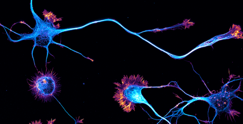
In addition to neurophysiological recording from S1 neurons, we also stained the cerebral cortex of one healthy control rat and one rat 3 days post-impact for c-Fos gene expression (Figure 4). As Figure 4 shows, in the post-concussion cortex, there is a visible and dramatic increase in gene expression within deep cortical layers (layers 5 and 6), but little-to-no labeling in the upper and middle layers (layers 2-4).
Figure 4. c-Fos labeling of gene expression in the S 1 cortex . Note the prominent staining of neurons’ nuclei , almost exclusively in the deep cortical layers , in the rat that suffered brain trauma 3 days earlier , whereas the cortex of a control rat that had no head trauma in its recent history shows no c-Fos labeling at all.
As this prominent c-Fos gene expression was confined to the deep cortical layers of the post-impact cortex, we looked for neurophysiological differences between the deep and mid-upper cortical layers. As Figure 5 shows, we do find prominent differences in the magnitude of spontaneous activity between the deep and mid-upper layers 3 days post-impact. The head impact transiently elevates spontaneous activity in all cortical layers, but by the 3rd day after impact, spontaneous activity in the mid-upper layers has declined to below-normal levels, while spontaneous activity in the deep layers remains elevated. Furthermore, the second head impact again elevates spontaneous activity in the mid-upper layers, but greatly suppresses it (below the control state) in the deep layers.
Figure 5.S1 spontaneous activity in the mid-upper and deep cortical layers. Plotted is the resting firing rate averaged over all the recorded neurons in each group.
Discussion
In this study we focused on 3 neurophysiological metrics of the cortical state: spontaneous spike firing rate, stimulus-evoked spike firing response, and inter-trial variability in stimulus-evoked responses. Spontaneous spike firing activity of the recorded neuronal population, in the absence of any stimulation, can be viewed as an indicator of the resting state of that neuronal population. The overall mean firing rate response of a cortical macrocolumn to vibrotactile stimulation of its RF center can be viewed as an indicator of responsivity of the recorded cortical columns to sensory stimulation in general. And inter-trial response variability averaged over a representative sample of neurons in the recorded macrocolumn can be viewed as an indicator of that macrocolumn’s reliability (or noisiness) in processing and transmitting sensory information.
At 6-12hr post-impact, the most prominent effect is on the spontaneous activity, which is elevated by 40%. In contrast, the effect on stimulus responsivity is minor (not rising to statistical significance). However, by the time we record from S1 again, 3 days later, in the mid-upper layers this initial boost has disappeared, and spontaneous activity greatly subsides (down to 65% of control level), although spontaneous activity remains high in the deep layers. Also, both stimulus responsivity and its inter-trial variability decline relative to their levels 6-12hr post-impact. Similar reductions in stimulus responsivity have been observed in rat S1 barrel cortex 24hr post-impact (Alwis et al., 2012; Johnstone et al., 2013). We also find c-Fos, an immediate early gene activated in neural tissues undergoing plastic changes, to be greatly elevated in the deep cortical layers and almost entirely unchanged elsewhere. This layer-specific c-Fos activation appears to be dependent on the nature of the brain injury, since Hall and Lifshitz (2010) did not see it in rat S1 in their midline fluid percussion brain injury model. As the axons of neurons in deep layers extend subcortically, travelling in the white matter for long distances, they should be more vulnerable than neurons in the mid-upper layers to shearing forces generated by weight-drop impact-induced acceleration/deceleration. It is likely that c-Fos activation and elevated spontaneous activity in the deep layer neurons reflect their response to shearing trauma to their axons.
It is known that shortly after a concussion the brain is in a vulnerable state and a second concussion in this period can cause far more severe damage. Indeed, we find that a second head impact 3 days after the first causes the S1 response recorded 6-12hr later to differ significantly from the response to the first impact. Most dramatic is the sharp decrease in spontaneous activity in deep cortical layers, instead of the small increase seen from a single impact. Less prominent, but still statistically significant, is the reduction of both the stimulus responsivity and its inter-trial variability, as compared to the first impact. Overall, the S1 effects of the second impact were nonlinear; the 6-12hr post-2nd impact group did not merely have the effects of an additional single impact ‘added on’ to the 6-12hr post-1st impact group. Indeed, using three basic measures of cortical functional state (mean spontaneous activity, variability of spontaneous activity, and mean stimulus response), it is possible to easily differentiate the four studied groups (Figure 6). The dramatic changes in both position and direction make it clear that the second impact is changing neural activity in different ways than the first.
In conclusion, our neurophysiological study of rats subjected to mild blunt head trauma demonstrates that mTBI does alter functionality of local neural mechanisms in the primary somatosensory cortex in a time-dependent manner.
Figure 6.The three brain-injured states and healthy controls are plotted in 3-D space defined by: (1) mean spontaneous activity of S1; (2) diversity of spontaneous activity levels among S1 neurons; and (3) mean firing rate of S1 macrocolumns in response to vibrotactile stimulation. The size of each ellipsoid corresponds to the standard error of the mean along each plotted dimension.
REFERENCES
Alwis, D. S., Yan, E. B., Morganti-Kossmann, M.-C., & Rajan, R. (2012). Sensory Cortex Underpinnings of Traumatic Brain Injury Deficits. PLoS ONE, 7(12), e52169. https://doi.org/10.1371/journal.pone.0052169
Carron, S. F., Alwis, D. S., & Rajan, R. (2016). Traumatic Brain Injury and Neuronal Functionality Changes in Sensory Cortex. Frontiers in Systems Neuroscience, 10. https://doi.org/10.3389/fnsys.2016.00047
Cole, W. R., Gregory, E., Arrieux, J. P., & Haran, F. J. (2017). Intraindividual Cognitive Variability: An Examination of ANAM4 TBI-MIL Simple Reaction Time Data from Service Members with and without Mild Traumatic Brain Injury. Journal of the International Neuropsychological Society, 24(2), 156–162. https://doi.org/10.1017/s1355617717001187
Favorov, O. V, Francisco, E., Holden, J., Kursun, O., Zai, L., & Tommerdahl, M. (2019). Quantification of Mild Traumatic Brain Injury via Cortical Metrics: Analytical Methods. Military Medicine, 184(Supplement_1), 228–236. https://doi.org/10.1093/milmed/usy411
Hall, K. D., & Lifshitz, J. (2010). Diffuse traumatic brain injury initially attenuates and later expands activation of the rat somatosensory whisker circuit concomitant with neuroplastic responses. Brain Research, 1323, 161–173. https://doi.org/10.1016/j.brainres.2010.01.067
Johnstone, V. P. A., Shultz, S. R., Yan, E. B., O’Brien, T. J., & Rajan, R. (2014). The Acute Phase of Mild Traumatic Brain Injury Is Characterized by a Distance-Dependent Neuronal Hypoactivity. Journal of Neurotrauma, 31(22), 1881–1895. https://doi.org/10.1089/neu.2014.3343
Johnstone, V. P. A., Yan, E. B., Alwis, D. S., & Rajan, R. (2013). Cortical Hypoexcitation Defines Neuronal Responses in the Immediate Aftermath of Traumatic Brain Injury. PLoS ONE, 8(5), e63454. https://doi.org/10.1371/journal.pone.0063454
Kane, M. J., Angoa-Pérez, M., Briggs, D. I., Viano, D. C., Kreipke, C. W., & Kuhn, D. M. (2012). A mouse model of human repetitive mild traumatic brain injury. Journal of Neuroscience Methods, 203(1), 41–49. https://doi.org/10.1016/j.jneumeth.2011.09.003
King, D. A., Hume, P. A., & Tommerdahl, M. (2018). Use of the Brain-Gauge Somatosensory Assessment for Monitoring Recovery from Concussion: A Case Study. In J Physiother Res (Vol. 2, Issue 1). iMedPub. www.corticalmetrics.com
Mychasiuk, R., Farran, A., Angoa-Perez, M., Briggs, D., Kuhn, D., & Esser, M. J. (2014). A Novel Model of Mild Traumatic Brain Injury for Juvenile Rats. Journal of Visualized Experiments, 94. https://doi.org/10.3791/51820
Mychasiuk, R., Farran, A., & Esser, M. J. (2014). Assessment of an Experimental Rodent Model of Pediatric Mild Traumatic Brain Injury. Journal of Neurotrauma, 31(8), 749–757. https://doi.org/10.1089/neu.2013.3132
Pearce, A. J., Kidgell, D. J., Frazer, A. K., King, D. A., Buckland, M. E., & Tommerdahl, M. (2019). Corticomotor correlates of somatosensory reaction time and variability in individuals with post concussion symptoms. , 37(1), 14–21. https://doi.org/10.1080/08990220.2019.1699045
Pearce, A. J., Tommerdahl, M., & King, D. A. (2019). Neurophysiological abnormalities in individuals with persistent post-concussion symptoms. Neuroscience, 408, 272–281. https://doi.org/10.1016/j.neuroscience.2019.04.019
Prins, M. L., Alexander, D., Giza, C. C., & Hovda, D. A. (2013). Repeated Mild Traumatic Brain Injury: Mechanisms of Cerebral Vulnerability. Journal of Neurotrauma, 30(1), 30–38. https://doi.org/10.1089/neu.2012.2399
Schiene, K., Bruehl, C., Zilles, K., Qu, M., Hagemann, G., Kraemer, M., & Witte, O. W. (1996). Neuronal Hyperexcitability and Reduction of GABAA-Receptor Expression in the Surround of Cerebral Photothrombosis. , 16(5), 906–914. https://doi.org/10.1097/00004647-199609000-00014
Tommerdahl, M., Dennis, R. G., Francisco, E. M., Holden, J. K., Nguyen, R., & Favorov, O. V. (2016). Neurosensory Assessments of Concussion. Military Medicine, 181(5S), 45–50. https://doi.org/10.7205/milmed-d-15-00172
Vagnozzi, R., Tavazzi, B., Signoretti, S., Amorini, A. M., Belli, A., Cimatti, M., Delfini, R., Di Pietro, V., Finocchiaro, A., & Lazzarino, G. (2007). TEMPORAL WINDOW OF METABOLIC BRAIN VULNERABILITY TO CONCUSSIONS. Neurosurgery, 61(2), 379–389. https://doi.org/10.1227/01.neu.0000280002.41696.d8
Waters, R., Li, C., & McCandlish, C. (1995). Relationship between the organization of the forepaw barrel subfield and the representation of the forepaw in layer IV of rat somatosensory cortex. Experimental Brain Research, 103(2). https://doi.org/10.1007/bf00231705
References
- Alwis, D. S., Yan, E. B., Morganti-Kossmann, M.-C., & Rajan, R. (2012). Sensory Cortex Underpinnings of Traumatic Brain Injury Deficits. PLoS ONE, 7(12), e52169. https://doi.org/10.1371/journal.pone.0052169
- Carron, S. F., Alwis, D. S., & Rajan, R. (2016). Traumatic Brain Injury and Neuronal Functionality Changes in Sensory Cortex. Frontiers in Systems Neuroscience, 10. https://doi.org/10.3389/fnsys.2016.00047
- Cole, W. R., Gregory, E., Arrieux, J. P., & Haran, F. J. (2017). Intraindividual Cognitive Variability: An Examination of ANAM4 TBI-MIL Simple Reaction Time Data from Service Members with and without Mild Traumatic Brain Injury. Journal of the International Neuropsychological Society, 24(2), 156–162. https://doi.org/10.1017/s1355617717001187
- Favorov, O. V, Francisco, E., Holden, J., Kursun, O., Zai, L., & Tommerdahl, M. (2019). Quantification of Mild Traumatic Brain Injury via Cortical Metrics: Analytical Methods. Military Medicine, 184(Supplement_1), 228–236. https://doi.org/10.1093/milmed/usy411
- Hall, K. D., & Lifshitz, J. (2010). Diffuse traumatic brain injury initially attenuates and later expands activation of the rat somatosensory whisker circuit concomitant with neuroplastic responses. Brain Research, 1323, 161–173. https://doi.org/10.1016/j.brainres.2010.01.067
- Johnstone, V. P. A., Shultz, S. R., Yan, E. B., O’Brien, T. J., & Rajan, R. (2014). The Acute Phase of Mild Traumatic Brain Injury Is Characterized by a Distance-Dependent Neuronal Hypoactivity. Journal of Neurotrauma, 31(22), 1881–1895. https://doi.org/10.1089/neu.2014.3343
- Johnstone, V. P. A., Yan, E. B., Alwis, D. S., & Rajan, R. (2013). Cortical Hypoexcitation Defines Neuronal Responses in the Immediate Aftermath of Traumatic Brain Injury. PLoS ONE, 8(5), e63454. https://doi.org/10.1371/journal.pone.0063454
- Kane, M. J., Angoa-Pérez, M., Briggs, D. I., Viano, D. C., Kreipke, C. W., & Kuhn, D. M. (2012). A mouse model of human repetitive mild traumatic brain injury. Journal of Neuroscience Methods, 203(1), 41–49. https://doi.org/10.1016/j.jneumeth.2011.09.003
- King, D. A., Hume, P. A., & Tommerdahl, M. (2018). Use of the Brain-Gauge Somatosensory Assessment for Monitoring Recovery from Concussion: A Case Study. In J Physiother Res (Vol. 2, Issue 1). iMedPub. www.corticalmetrics.com
- Mychasiuk, R., Farran, A., Angoa-Perez, M., Briggs, D., Kuhn, D., & Esser, M. J. (2014). A Novel Model of Mild Traumatic Brain Injury for Juvenile Rats. Journal of Visualized Experiments, 94. https://doi.org/10.3791/51820
- Mychasiuk, R., Farran, A., & Esser, M. J. (2014). Assessment of an Experimental Rodent Model of Pediatric Mild Traumatic Brain Injury. Journal of Neurotrauma, 31(8), 749–757. https://doi.org/10.1089/neu.2013.3132
- Pearce, A. J., Kidgell, D. J., Frazer, A. K., King, D. A., Buckland, M. E., & Tommerdahl, M. (2019). Corticomotor correlates of somatosensory reaction time and variability in individuals with post concussion symptoms. Somatosensory & Motor Research, 37(1), 14–21. https://doi.org/10.1080/08990220.2019.1699045
- Pearce, A. J., Tommerdahl, M., & King, D. A. (2019). Neurophysiological abnormalities in individuals with persistent post-concussion symptoms. Neuroscience, 408, 272–281. https://doi.org/10.1016/j.neuroscience.2019.04.019
- Prins, M. L., Alexander, D., Giza, C. C., & Hovda, D. A. (2013). Repeated Mild Traumatic Brain Injury: Mechanisms of Cerebral Vulnerability. Journal of Neurotrauma, 30(1), 30–38. https://doi.org/10.1089/neu.2012.2399
- Schiene, K., Bruehl, C., Zilles, K., Qu, M., Hagemann, G., Kraemer, M., & Witte, O. W. (1996). Neuronal Hyperexcitability and Reduction of GABAA-Receptor Expression in the Surround of Cerebral Photothrombosis. Journal of Cerebral Blood Flow & Metabolism, 16(5), 906–914. https://doi.org/10.1097/00004647-199609000-00014
- Tommerdahl, M., Dennis, R. G., Francisco, E. M., Holden, J. K., Nguyen, R., & Favorov, O. V. (2016). Neurosensory Assessments of Concussion. Military Medicine, 181(5S), 45–50. https://doi.org/10.7205/milmed-d-15-00172
- Vagnozzi, R., Tavazzi, B., Signoretti, S., Amorini, A. M., Belli, A., Cimatti, M., Delfini, R., Di Pietro, V., Finocchiaro, A., & Lazzarino, G. (2007). TEMPORAL WINDOW OF METABOLIC BRAIN VULNERABILITY TO CONCUSSIONS. Neurosurgery, 61(2), 379–389. https://doi.org/10.1227/01.neu.0000280002.41696.d8
- Waters, R., Li, C., & McCandlish, C. (1995). Relationship between the organization of the forepaw barrel subfield and the representation of the forepaw in layer IV of rat somatosensory cortex. Experimental Brain Research, 103(2). https://doi.org/10.1007/bf00231705